Neuro Jamming
Modeling Non-Linear Dynamics of Complex Material in the Brain
Saif Haobsh - MIT: Principles of Neuroengineering
Introduction to Jamming
In Nature’s 1998 November Journal, a seminal paper titled “Nonlinear dynamics: Jamming is not just cool anymore” was published resurfacing the concepts behind the material dynamics of jammed-states. A broad class of materials including foams, glassy molecular systems, colloids, and granular materials are known to form jammed states. A jammed system can withstand small applied forces without deforming irreversibly, while unjammed systems flow freely.
The diagram in figure 1 elegantly relates the jamming transition in three axes: temperature, load, and inversely to density. For example, glass can be considered in a jammed state as a super-cooled liquid with super-high viscosity, and can be unjammed with changes in temperature, density, or applied load (See Figure 1).
This concept of non-linear properties of materials, blending the perception of phase-state and phase-change, might lend itself to understanding the complexity of the brain, with its largest merit consolidating multiple axes of material behavior under one rubric. Although temperature, density, and load serve as very limited variables in which the human brain can withstand without causing irreversible damage, the load/density plane provides a framework for which the neuro-jamming concept can be tested. Consequently, when applied load on the brain is typically explored in the context of causing traumatic brain injury (TBI), or irreversible deformation, neuro-jamming explores the context of reversible deformation. Essentially, this paper investigates the small threshold in which force, innocuously applied on the brain, can shed light on the brains highly undiscovered behaviors.
Brain Mechanics
The viscoelastic properties of the brain are essential in characterizing the mechanical deformation. In a paper titled “Viscoelastic properties of human cerebellum using magnetic resonance elastography” published in the Journal of Biomechanics in July 2011, the linear properties of the brain were first characterized through Magnetic Resonance Elastrography (MRE) . The findings of the paper suggested that regional differences existed in relation to the tissue type; “white matter was more viscous than grey matter and the cerebrum was more viscous than the cerebellum”. Although linear in nature, these findings were the first to document viscoelastic properties of the brain suggesting that different regions of brain tissue will possibly respond to mechanical loading differently.
Looking further into the Biomechanics of soft tissue, especially that of the human brain, it was previously shown that stress/strain curves for compression rates exhibited concavity suggesting highly non-linear properties (See Figure 2).
Consequently, nonlinear dynamics of brain tissue proves to be a more adequate basis for understanding and testing both theoretical and experimental models. In 2012, it was shown that dilatational waves, which develop into shock waves, attenuate at higher rates with increasing load, with localization of energy accumulations. These accumulations of high stress gradients can result in micro-scale tissue damages when elastic limits are breached.
Although forces on the body can be sourced from mechanical loading, the complimentary field of sonography, or imaging through sound, has proven to shed light on understanding shear wave. Experimentally, forces on the brain needed for shear wave propagation have been explored from acoustical sources as well.
Particularly in the biomechanics of shear deformation and shock waves fluid dynamics and ultrasonography have direct application. Outlined in “Shear Shock Waves Observed in the Brain” , an experimental setup involving a porcine ex-vivo brain in a vat of gelatin with displacement produced by an electromechanical shaker. An ultrasound transducer is used to read wave propagation. The high-frame rate ultrasound technique showed that nonlinear dynamics of shear shock waves, with relatively short wavelength was sufficient for measurable readings (See Figure 3). More importantly the mechanism was consistent with theoretical predictions of nonlinearities.
In the context of Traumatic Brain Injury (TBI), a pathology with extreme urgency as the leading cause of accident related death and disability worldwide, wave propagation has previously been studied as an impetus for causing TBI. The concepts that follow “Neuro-Jamming” however have potential to channel applied force as an active method of modeling and imaging previous deformation in the brain.
Modeling the Brain
Modeling the brain has played an integral part of investigating TBI and external loading. The subarachnoid space (SAS), was presented as the critical interface between the skull and the brain, as a damping and moving interstitial zone. A paper put forth in 2011 by Saboori and Sadegh for Scientia Iranica laid out three material models for optimal properties; these include soft solid material, viscous fluid, and porous media. The findings placed all three models as suitable values for modeling and FE analyses.
The following is a table of material properties used for mechanical simulations based on findings in “Material modeling of the head’s subarachnoid space”, which consolidates from previous literature on non-linear viscoelastic properties of the brain.
Including the material properties layed out for the model, a simplified massing model of the head was developed, containing roughly offset layers of “scalp, skull, dura mater, SAS, white matter and grey matter” (See Figure 4). Although the massing model provides a helpful framework for understanding the regions of the varying properties, the simplified model is too reductive for critical nuances.
Voxel-Based Modeling
This proposal seeks to put forth a method for modeling mass and material attributed with higher granularity. With recent breakthroughs in additive material manufacturing, emergent tools in voxel-based modeling provide added value. Volumetric, multi-material, and computational in nature, these tools may provide nuanced considerations necessary. Monolith, software developed by Panagiotis Michalatos and Andrew Payne, will be used for the purposes of this proposal.
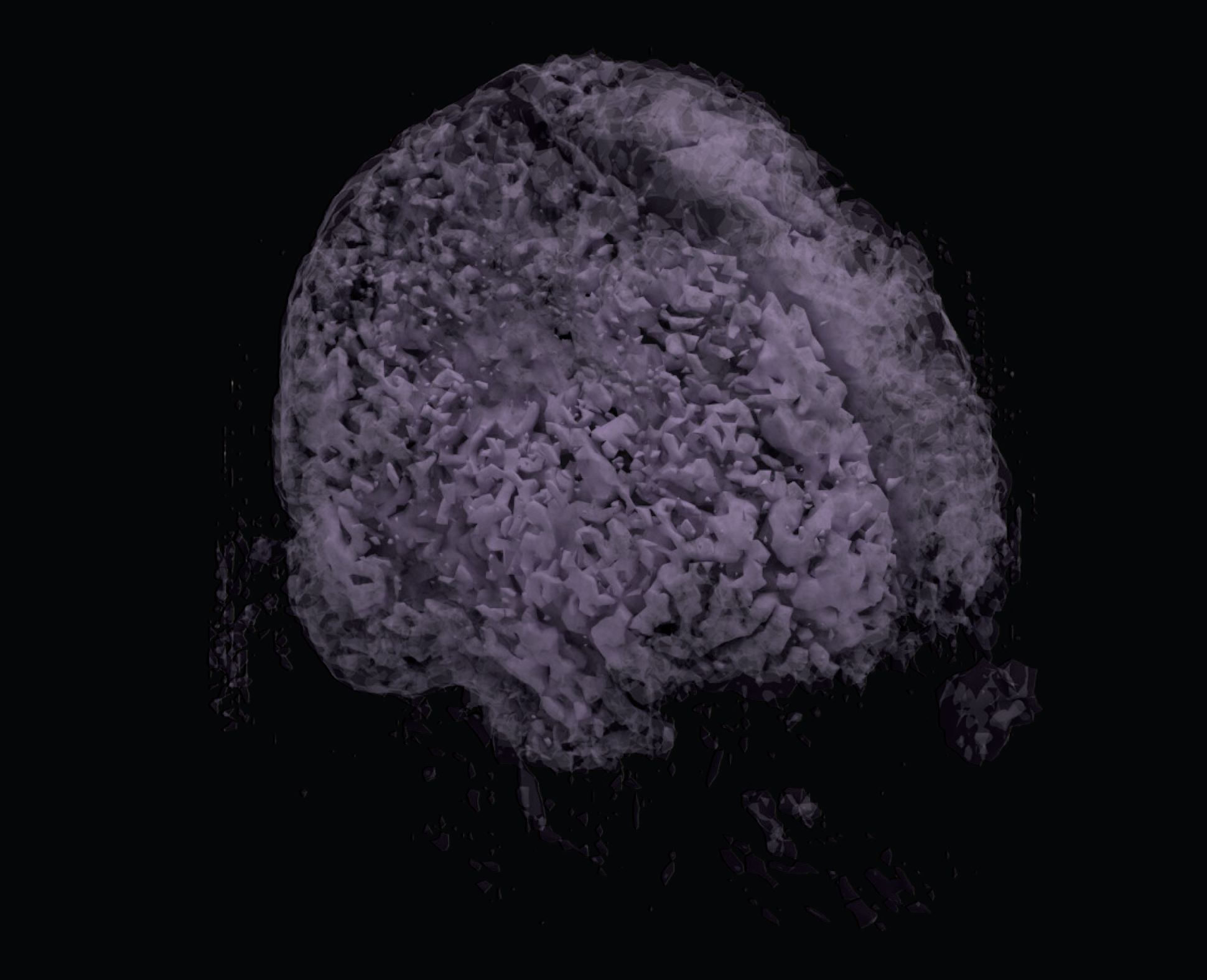
Image Stack of Volume
The first step is to obtain a voxel image of the three dimensional volume with channels. For the purposed of this proposed, an fMRI image of stack of DICOM files in greyscale thresholds will serve as the voxel channel normalized to [0.0 to 1.0]. The compositions of the layers form a field of boxes that each carry individual packets information, i.e. material properties (See Figure 5).
Isosurface Extrapolation
From the voxel channel, isovalues representing the threshold within a field define topologies of corresponding surfaces. These isovalues can represent solid-void or material transitions . From the range [0.0 to 1.0], isovalues are extracted in between in equal intervals (See Figure 6).
Topology Optimization Analysis
Topology optimization is structural analysis which optimizes material distributions within a volume given applied loads and boundary conditions. In this demonstration, a topology optimization analysis tool available from within Monolith is being used. The setup includes the iso-value thresholds of tissue densities, applied loads, boundary conditions, and mechanical properties of materials. Stress lines and mesh deformations are visualized as outputs from the analysis (See Figure 7 and Figure 8).
Applications and Need
The findings in this proposal present two tracks for which potential application of the technology exists. The first of which would find use in post traumatic brain injury scenarios. Irreversible damage caused by blast loading to the head can be simulated recreating the force loads on the brain both through speculative and empirical data collected from accident site and in-vivo imaging. The stress lines obtained through the topology optimization analysis can provide a framework for surgical strategy in healthcare administration. Essentially, information of damage not-obtainable through existing imaging techniques, can be extrapolated through simulation for medical intervention. Providing a data driven model of mechanical deformation can potentially reduce further damage of invasive surgical procedures.
The second potential application is of obtaining longitudinal models of tissue deformation in post trauma events with innocuous micro impulses. Electromechanical shaking during imaging may provide deeper insight into local maxima of deformation in tissue, not perceivable while patient is in equilibrium. With the a priori consideration of the brain to be in a jammed state, Neuro-Jamming provides potential value in reframing our understanding of the mysteries of our brain.
Bibliography
Dogan, Firat, and M. Serdar Celebi. “Real-Time Deformation Simulation of Non-Linear Viscoelastic Soft Tissues.” Simulation, vol. 87, no. 3, Aug. 2010, pp. 179–187., doi:10.1177/0037549710364532.
Espindola, David, and Gianmarco Pinton. “Shear Shock Waves Observed in the Ex-Vivo Brain.” 2017 IEEE International Ultrasonics Symposium (IUS), 2017, doi:10.1109/ultsym.2017.8092901.
Haut, Roger C. “Biomechanics of Soft Tissue.” Accidental Injury, 2002, pp. 228–253., doi:10.1007/978-0-387-21787-1_11.
Laksari, K. (2013). Nonlinear viscoelastic wave propagation in brain tissue (Order No. 3611150). Available from ProQuest Dissertations & Theses Global. (1501656347). Retrieved from http://search.proquest.com.ezp-prod1.hul.harvard.edu/docview/1501656347?accountid=11311
Laksari, Kaveh, et al. “Computational Simulation of the Mechanical Response of Brain Tissue under Blast Loading.” Biomechanics and Modeling in Mechanobiology, vol. 14, no. 3, Oct. 2014, pp. 459–472., doi:10.1007/s10237-014-0616-2.
Liu, Andrea J., and Sidney R. Nagel. “Jamming Is Not Just Cool Any More.” Nature, vol. 396, no. 6706, 1998, pp. 21–22., doi:10.1038/23819.
Saboori, P., and A. Sadegh. “Material Modeling of the Head’s Subarachnoid Space.” Scientia Iranica, vol. 18, no. 6, 2011, pp. 1492–1499., doi:10.1016/j.scient.2011.11.032.
Toledo, Esteban, et al. “The Young Brain and Concussion: Imaging as a Biomarker for Diagnosis and Prognosis.” Neuroscience & Biobehavioral Reviews, vol. 36, no. 6, 2012, pp. 1510–1531., doi:10.1016/j.neubiorev.2012.03.007.
Iso-surface topologies of brain matter
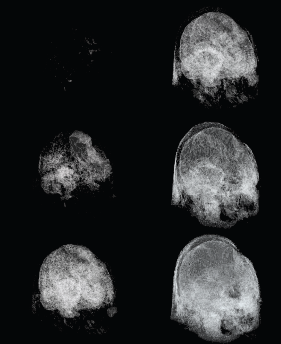
Figure 5: Image Stack of Volumes
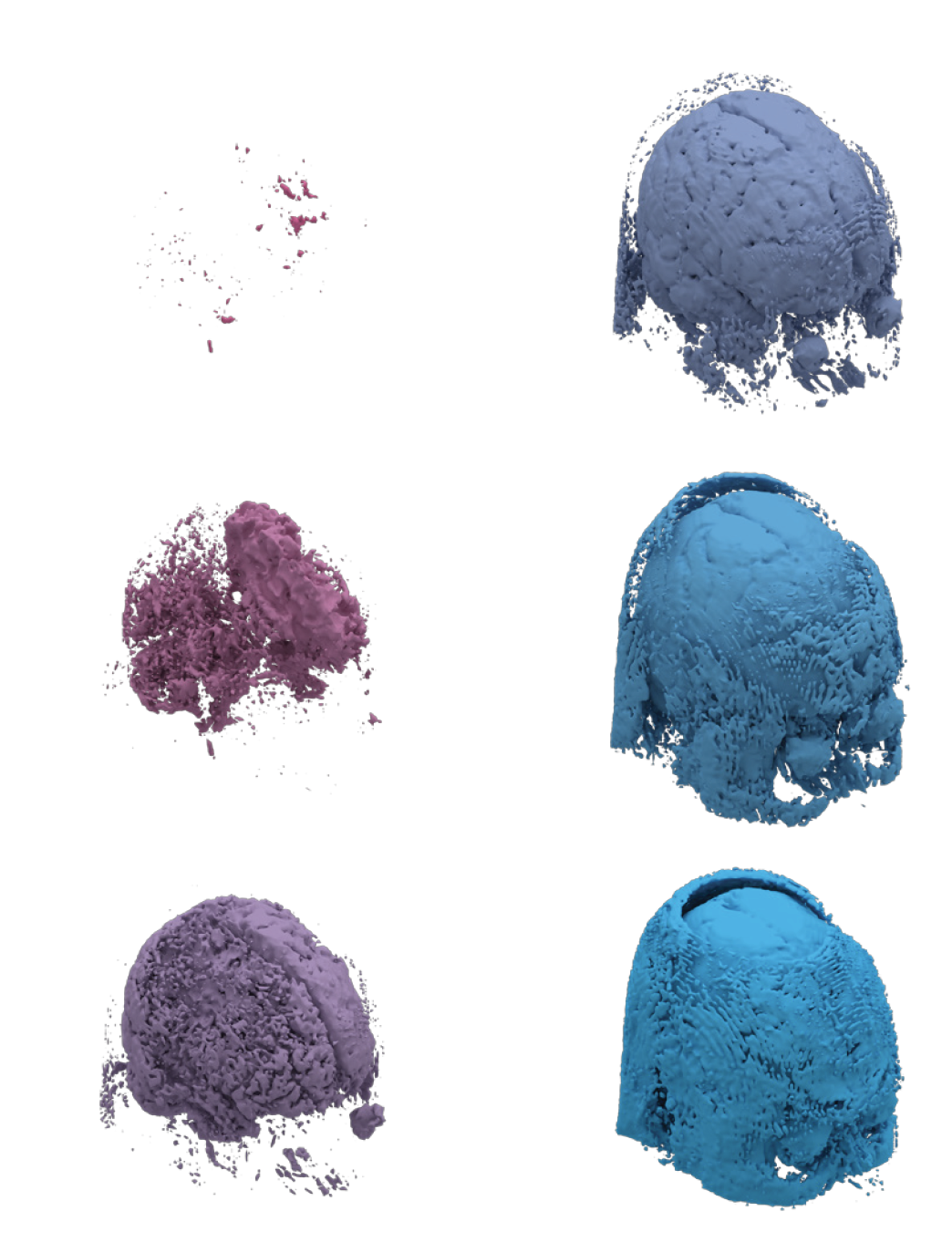
Figure 6: Isosurface Topologies at varying thresholds.
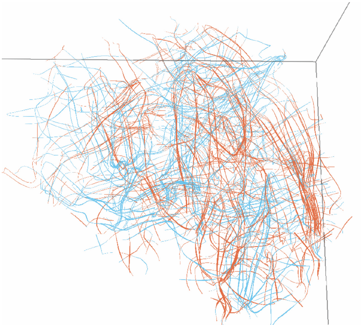
Figure 7: Stress Line Visualization
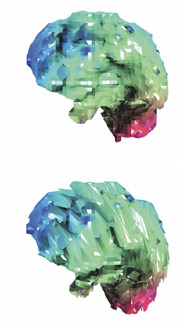
Figure 8: Augmented mesh deformation before (top) and after (bottom) loading.